Summary:
UCLA researchers in the Department of Bioengineering have developed a novel fluorescence lifetime imaging microscopy technique which enables fast 3D imaging using low dimensional detectors.
Background:
Fluorescence lifetime imaging microscopy, or FLIM, measures the fluorescence lifetime of probes embedded in tissue samples which enables visualization of biological samples at the molecular level. However, traditional FLIM methods suffer from long acquisition time, especially in 3D imaging as slices must be taken at many points along the depth axis. Other technologies such as Single-photon avalanche diodes (SPADs) utilize conventional gated cameras in both temporal resolution and sensitivity, but significant amounts of information in each pixel is lost to complicated processing requirements of SPAD arrays. Linear SPAD arrays allow for much more useful information in each pixel, increasing image quality, but 3D imaging using linear SPAD arrays greatly increases acquisition time. There is a clear need for a method allowing linear SPAD arrays to image 3D samples with fast acquisition times using low-cost detectors.
Innovation:
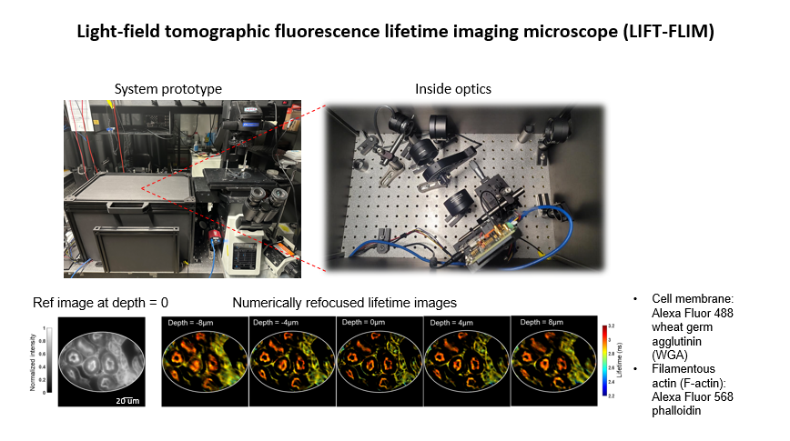
UCLA researchers led by Dr. Liang Gao have advanced a computational imaging method, light-field tomographic fluorescence lifetime imaging microscopy, LIFT-FLIM, which enables use of low dimensional detectors for high dimensional imaging. The method transforms volumetric images into lines which can then be recorded by linear SPAD cameras, which are lower cost and more accessible to general research labs. This technique shows unparalleled single-photon sensitivity and post-processing time of less than 0.3 seconds.
Credit: Liang Gao
Potential Applications:
- Basic research
- Translational research
- Organoid imaging
- Biological and clinical samples
Advantages:
- 3D imaging using low-cost detectors
- Fast processing time
- High pixel fill factor
- Compatible with spectral FLIM
Development-To-Date:
Technique has been validated using standardized samples and demonstrated on samples of biological tissues
Related Papers:
Liang Gao et. al, Light-field tomographic fluorescence lifetime imaging microscopy. Preprint https://doi.org/10.21203/rs.3.rs-2883279/v1.
Reference:
UCLA Case No. 2023-130
Lead Inventor:
Liang Gao